
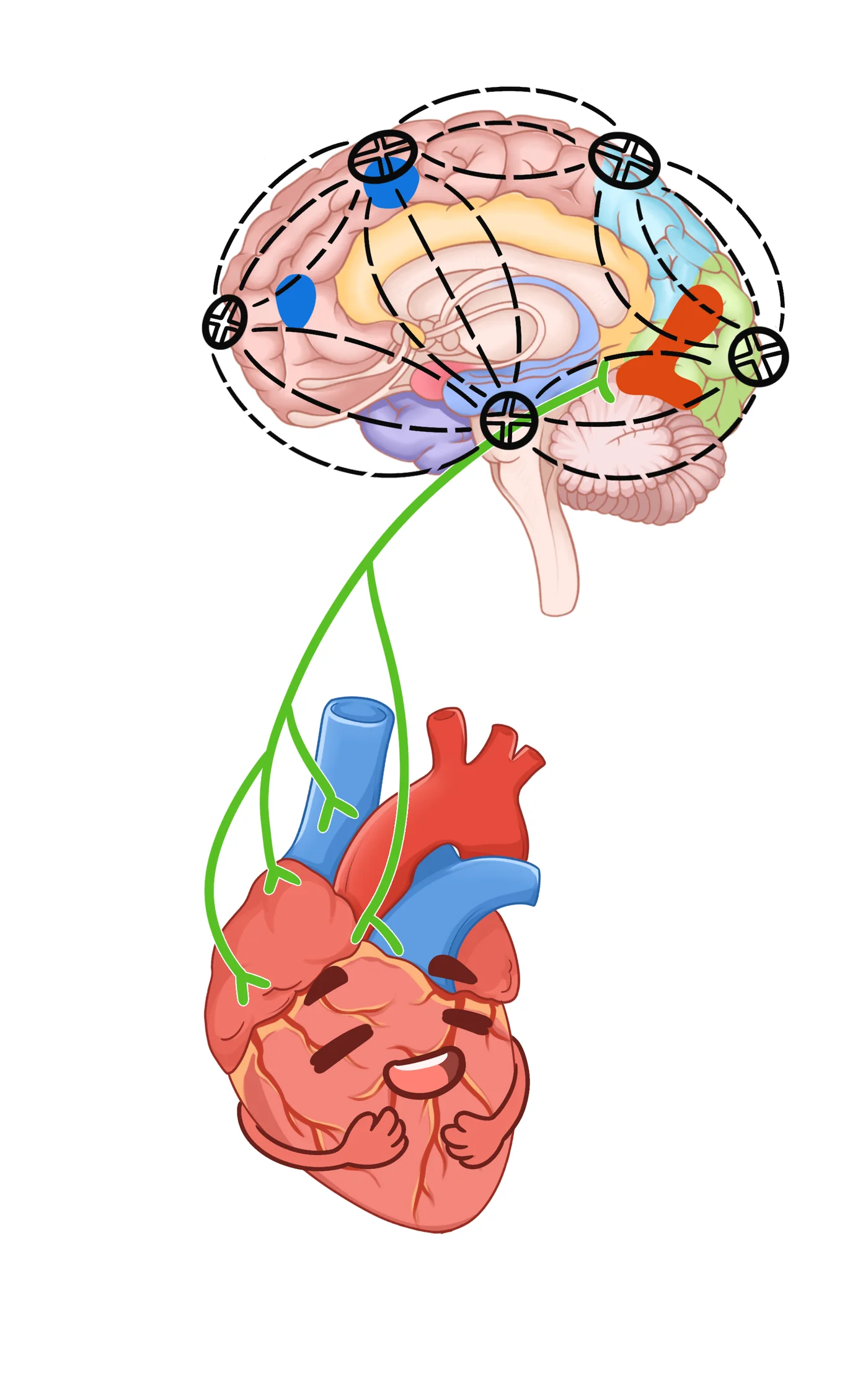
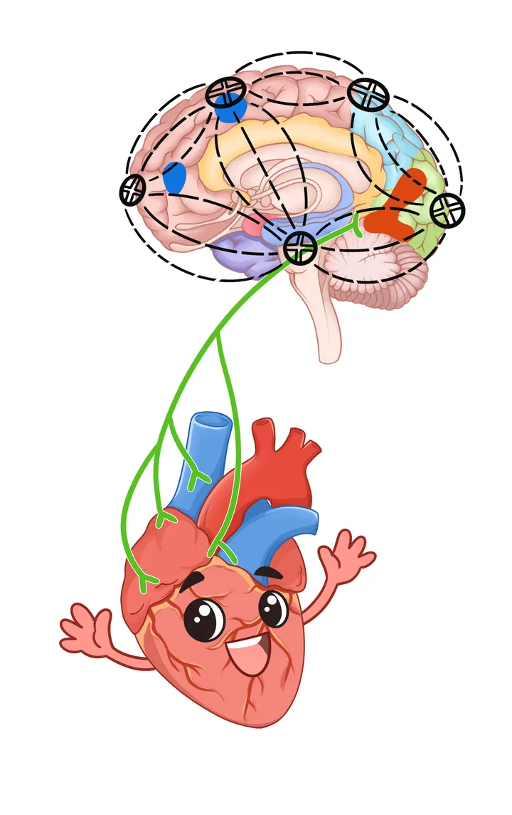
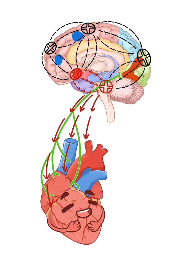
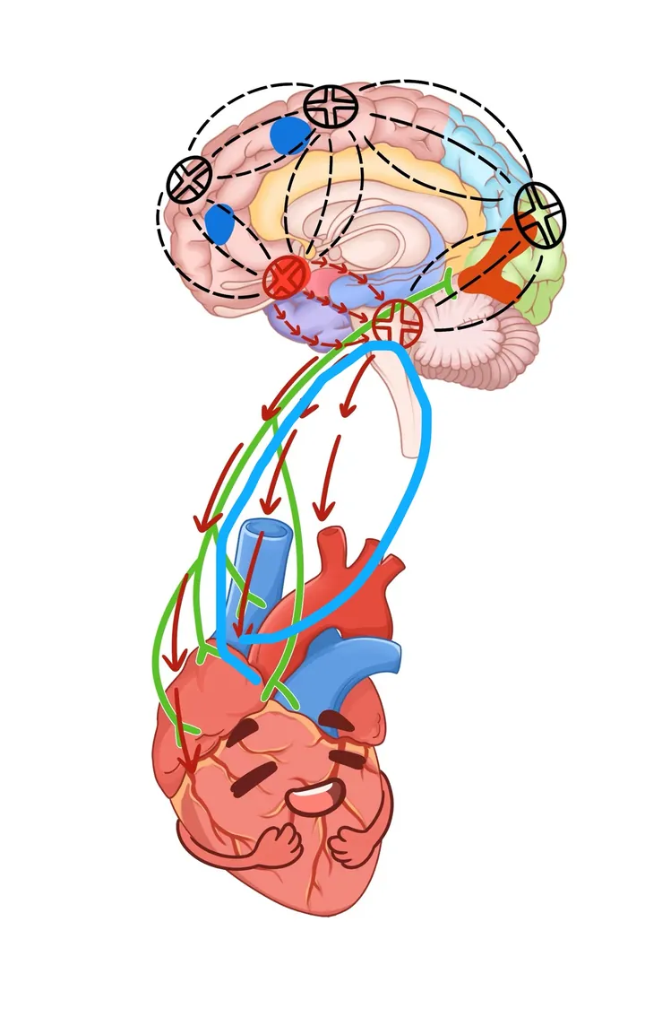
畫面上下排布大腦與心臟,中間以連線示意腦與心之間的雙向聯繫;大腦以功能分區着色,心臟被賦予笑臉。這種擬人化處理讓“腦—心軸”概念變得可親近。四個構圖高度相近的版本同作呈現,以存檔版本分別展示,不推定其中某一版爲最終稿。
圖背後的故事
引言:睡眠剝奪(SD)顯著擾亂心腦軸的穩態,而深部磁刺激(DMS)作爲一種無創、安全的方法,其神經調控作用仍不清楚。方法:招募 60 名健康成年男性進行 36 小時睡眠剝奪研究,按個人意願分入 DMS 組或對照組;所有人在實驗起點與終點接受心音測量和功能磁共振成像掃描。恢復睡眠階段,DMS 組在入睡前與醒來時各接受一次 30 分鐘深部磁刺激。研究用雙因素分析揭示狀態變化與組間干預效應,用 Spearman 秩相關評估腦活動與心活動的關聯,用線性迴歸探索 DMS 經腦區調節心活動的效應,並用自助法(bootstrap)檢驗中介效應。結果:DMS 組心動週期時長爲 0.81±0.04 秒,對照組爲 0.80±0.03 秒,DMS 呈延長效應(F=0.32,p=0.02),且 DMS 組各項心率與強度指標值均低於對照組(p<0.01)。雙因素分析顯示左側腦島與眶額下回差異顯著,DMS 後其 DC_Weight(0.25)值分別低於 0.50(p<0.01)與 0.42(p<0.01)。相關分析證實左眶額下回、左腦島與心音指標呈負相關(p<0.05):Δ左眶額下回與 Δ收縮期強度(rho=−0.33)、Δ舒張期強度(rho=−0.41)、ΔS1 強度(rho=−0.36)、ΔS2 強度(rho=−0.43)負相關;Δ左腦島與 Δ舒張期強度(rho=−0.36)、ΔS1 強度(rho=−0.33)、ΔS2 強度(rho=−0.36)負相關。中介效應分析顯示 DMS 通過干預腦區影響 S2 強度。結論:以上發現提示 36 小時睡眠剝奪後心腦軸存在因果效應;DMS 的無創干預可有效調節睡眠剝奪後的腦與心功能、促進穩態平衡。DMS 能夠影響心腦軸,爲長時間睡眠剝奪後的恢復提供了一種手段。
補充:作品創作背景
腦和心不是各自運轉的機器:壓力、睡眠、情緒都會通過神經與內分泌通路影響心臟。相關研究從兩個方向展開——急性睡眠剝奪下腦功能與心音的變化,以及深部磁刺激對腦心軸影響的探索性研究(見側欄)。
本圖爲機制示意,省略大量解剖細節,不能作爲醫學診斷或解剖教學依據。
論文證據
Figure 8 · derivative(同底稿的論文編輯版)
The effect of deep magnetic stimulation on the cardiac-brain axis post-sleep deprivation: a pilot study
論文摘要譯文(本站譯,忠實原文)
引言:睡眠剝奪(SD)顯著擾亂心腦軸的穩態,而深部磁刺激(DMS)作爲一種無創、安全的方法,其神經調控作用仍不清楚。方法:招募 60 名健康成年男性進行 36 小時睡眠剝奪研究,按個人意願分入 DMS 組或對照組;所有人在實驗起點與終點接受心音測量和功能磁共振成像掃描。恢復睡眠階段,DMS 組在入睡前與醒來時各接受一次 30 分鐘深部磁刺激。研究用雙因素分析揭示狀態變化與組間干預效應,用 Spearman 秩相關評估腦活動與心活動的關聯,用線性迴歸探索 DMS 經腦區調節心活動的效應,並用自助法(bootstrap)檢驗中介效應。結果:DMS 組心動週期時長爲 0.81±0.04 秒,對照組爲 0.80±0.03 秒,DMS 呈延長效應(F=0.32,p=0.02),且 DMS 組各項心率與強度指標值均低於對照組(p<0.01)。雙因素分析顯示左側腦島與眶額下回差異顯著,DMS 後其 DC_Weight(0.25)值分別低於 0.50(p<0.01)與 0.42(p<0.01)。相關分析證實左眶額下回、左腦島與心音指標呈負相關(p<0.05):Δ左眶額下回與 Δ收縮期強度(rho=−0.33)、Δ舒張期強度(rho=−0.41)、ΔS1 強度(rho=−0.36)、ΔS2 強度(rho=−0.43)負相關;Δ左腦島與 Δ舒張期強度(rho=−0.36)、ΔS1 強度(rho=−0.33)、ΔS2 強度(rho=−0.36)負相關。中介效應分析顯示 DMS 通過干預腦區影響 S2 強度。結論:以上發現提示 36 小時睡眠剝奪後心腦軸存在因果效應;DMS 的無創干預可有效調節睡眠剝奪後的腦與心功能、促進穩態平衡。DMS 能夠影響心腦軸,爲長時間睡眠剝奪後的恢復提供了一種手段。
文章摘要(中文譯文 · 原文照錄)
中文譯文 引言 睡眠剝奪(SD)會顯著破壞心腦軸的穩態,而深部磁刺激(DMS)這一無創、安全的神經調控方法對其影響仍知之甚少。方法 研究招募60名健康成年男性進行36小時睡眠剝奪,按個人意願分配至DMS組或對照組;所有受試者在實驗起點與終點接受心音測量和功能磁共振成像(fMRI)掃描。恢復睡眠階段,DMS組在入睡前與覺醒時各接受一次30分鐘刺激。採用雙因素分析揭示兩組在兩種狀態下的變化與干預效應,以Spearman等級相關分析評估腦活動與心臟活動的相關性,用線性迴歸分析探討DMS對調控心臟活動的腦區的影響,並以自助法(bootstrapping)驗證中介效應。結果 DMS組心動週期時長爲0.81 ± 0.04 s,對照組爲0.80 ± 0.03 s,DMS呈延長效應(F = 0.32,p = 0.02),且各項心音頻率與強度指標均低於對照組(p < 0.01)。双因素分析显示左侧脑岛与眶部额下回差异显著,DMS后 DC_Weight(0.25)值分别降低0.50(p < 0.01)与0.42(p < 0.01)。相关分析证实左侧眶部额下回、左侧脑岛与心音指标呈负相关(p < 0.05):Δ左侧眶部额下回与Δ收缩期强度(rho = −0.33)、Δ舒张期强度(rho = −0.41)、Δ S1强度(rho = −0.36)、Δ S2强度(rho = −0.43)负相关(均p < 0.05);Δ左侧脑岛与Δ舒张期强度(rho = −0.36)、Δ S1强度(rho = −0.33)、Δ S2强度(rho = −0.36)负相关。中介效应分析表明,DMS通过作用于脑区影响S2强度。结论 这些发现提示36小时睡眠剥夺后心脑轴存在因果效应;DMS无创干预可有效调节睡眠剥夺后的脑与心功能、促进稳态平衡,为长时间睡眠剥夺后的平衡恢复提供了一种手段。
Introduction Sleep deprivation (SD) significantly disrupts the homeostasis of the cardiac-brain axis, yet the neuromodulation effects of deep magnetic stimulation (DMS), a non-invasive and safe method, remain poorly understood. Methods Sixty healthy adult males were recruited for a 36-h SD study, they were assigned to the DMS group or the control group according to their individual willing. All individuals underwent heart sound measurements and functional magnetic resonance imaging scans at the experiment's onset and terminal points. During the recovery sleep phase, DMS was applied twice for 30 min before sleep onset and upon awakening to the individuals in the DMS group. Two-factor analysis was used to disclose the changes in two status and intervention effect in groups, along with Spearman rank correlation analysis to assess the correlation between brain activity and heart activity, the linear regression analysis was performed to explore the effect of DMS on brain regions to regulated the heart activity. Additionally, bootstrapping analysis was employed to verify the mediation effect. Results The results indicated that the DMS group cardiac cycle duration was 0.81 ± 0.04 s, CON group was 0.80 ± 0.03 s, DMS presented a prolong effect ( F = 0.32, p = 0.02), and all heart frequency and intensity indexes value were lower than CON group ( p < 0.01). Two-factor analysis demonstrated the significant differences in the left insula and orbitofrontal inferior gyrus, which DC_Weight (0.25) value were lower 0.50 ( p < 0.01), 0.42 ( p < 0.01) after DMS. Furthermore, the correlation analysis confirmed that the negative association between the left orbital inferior frontal and left insula with the heart sound index ( p < 0.05), such as Δ left orbital inferior frontal were negatively correlated with Δ Systolic_intensity (rho = −0.33, p < 0.05), Δ Diastolic_intensity (rho = −0.41, p < 0.05), Δ S1_intensity (rho = −0.36, p < 0.05), and Δ S2_intensity (rho = −0.43, p < 0.05). Δ Left insula was negatively correlated with Δ Diastolic_intensity (rho = −0.36, p < 0.05), Δ S1_intensity (rho = −0.33, p < 0.05), and Δ S2_intensity (rho = −0.36, p < 0.05). Mediated effect analysis showed that DMS affected S2_intensity by intervening in brain regions. Conclusion These findings suggest a causal effect on the cardiac-brain axis following 36 h of SD. The non-invasive intervention of DMS effectively regulates both brain and heart functions after SD, promoting homeostatic balance. The DMS can affect the cardiac-brain axis, offering a means to restore balance following extended periods of SD.
匹配說明:作品圖 F137(腦心軸擬人插畫)同一底稿的論文編輯版;腦區與擬人心臟一致,節點、箭頭與構圖有調整,見論文 Figure 8。
doi.org/10.3389/fnins.2024.1464299 · PubMed 39867450 · PMC 全文
題錄、日期與摘要據 PMC 原文;圖片再利用須遵守原文許可與署名要求。