
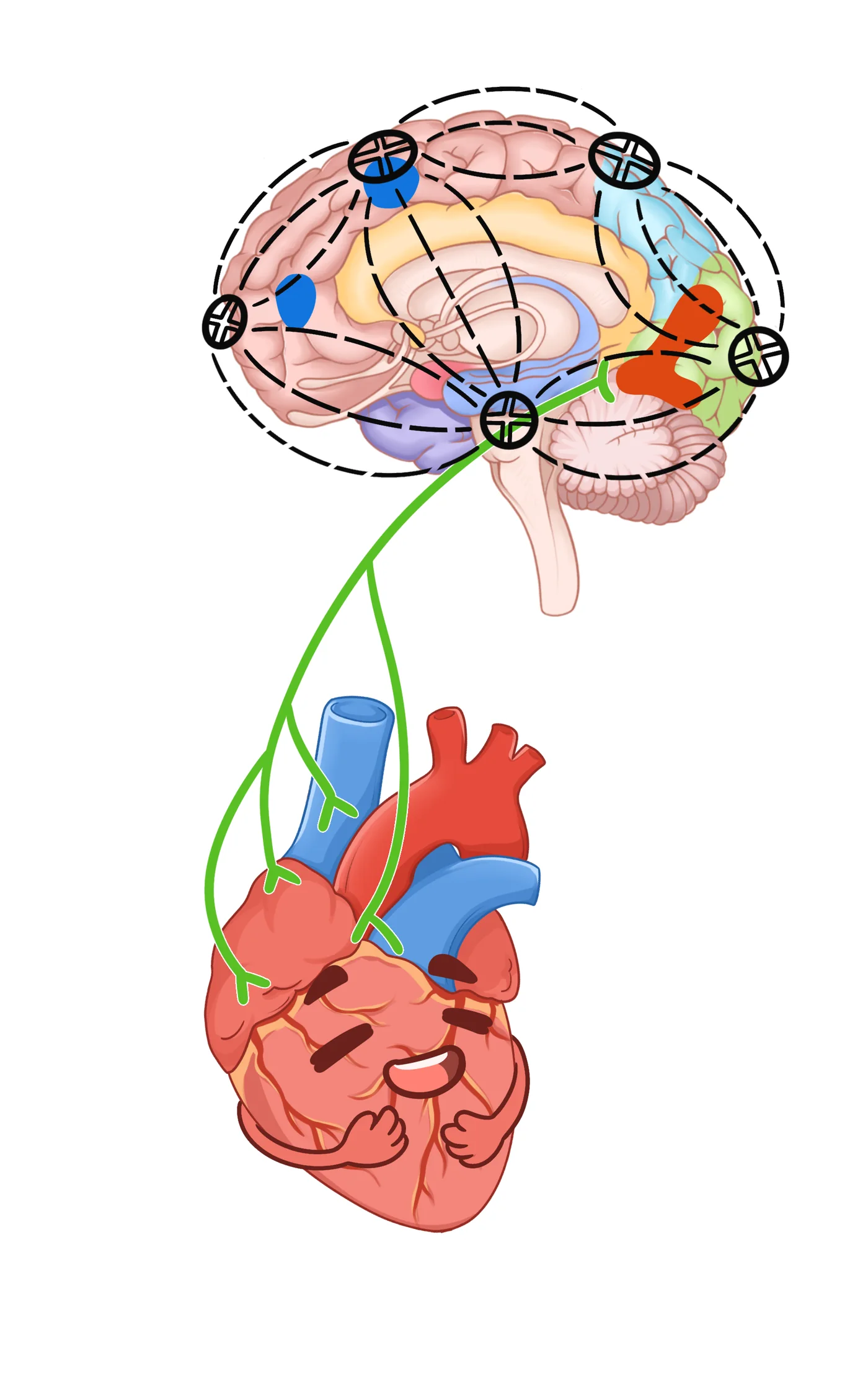
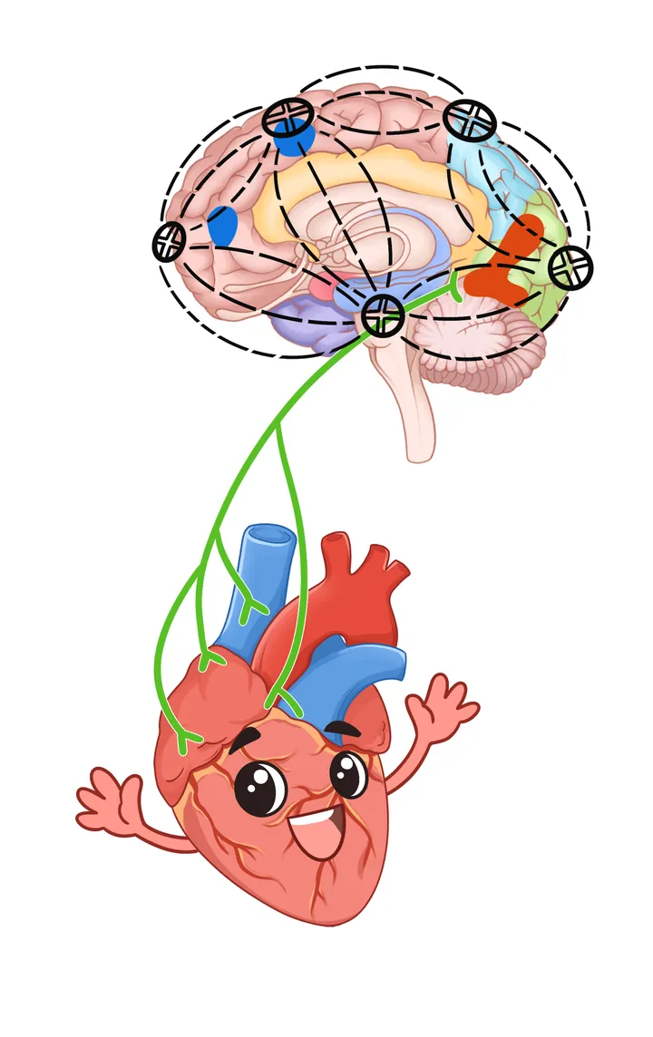
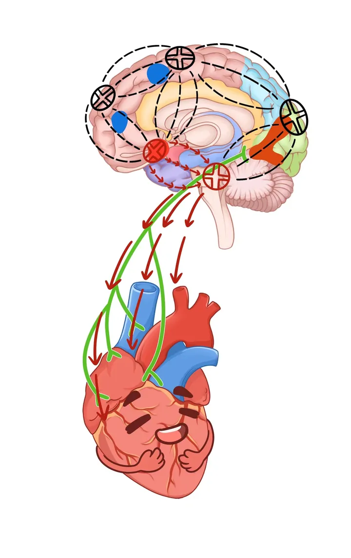
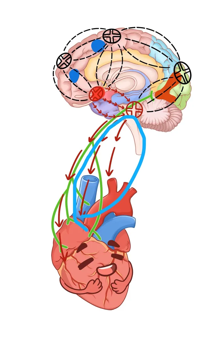
Brain above, heart below, connected by lines suggesting their two-way link; the brain is coloured by functional area and the heart given a smile. This personification makes the brain–heart axis approachable. Four highly similar compositions are shown as archived versions; none is assumed to be the final draft.
The Story Behind the Image
addendum: creation background
Brain and heart are not independent machines: stress, sleep and emotion all reach the heart via neural and endocrine pathways. Related research runs in two directions — brain and heart-sound changes under acute sleep deprivation, and an exploratory study of deep magnetic stimulation on the brain–heart axis (see sidebar).
This is a mechanism sketch; many anatomical details are omitted. It is not a basis for diagnosis or anatomy teaching.
Paper evidence
Figure 8 · derivative (paper-edited version of the same artwork)
The effect of deep magnetic stimulation on the cardiac-brain axis post-sleep deprivation: a pilot study
Article abstract (English original)
Introduction Sleep deprivation (SD) significantly disrupts the homeostasis of the cardiac-brain axis, yet the neuromodulation effects of deep magnetic stimulation (DMS), a non-invasive and safe method, remain poorly understood. Methods Sixty healthy adult males were recruited for a 36-h SD study, they were assigned to the DMS group or the control group according to their individual willing. All individuals underwent heart sound measurements and functional magnetic resonance imaging scans at the experiment's onset and terminal points. During the recovery sleep phase, DMS was applied twice for 30 min before sleep onset and upon awakening to the individuals in the DMS group. Two-factor analysis was used to disclose the changes in two status and intervention effect in groups, along with Spearman rank correlation analysis to assess the correlation between brain activity and heart activity, the linear regression analysis was performed to explore the effect of DMS on brain regions to regulated the heart activity. Additionally, bootstrapping analysis was employed to verify the mediation effect. Results The results indicated that the DMS group cardiac cycle duration was 0.81 ± 0.04 s, CON group was 0.80 ± 0.03 s, DMS presented a prolong effect ( F = 0.32, p = 0.02), and all heart frequency and intensity indexes value were lower than CON group ( p < 0.01). Two-factor analysis demonstrated the significant differences in the left insula and orbitofrontal inferior gyrus, which DC_Weight (0.25) value were lower 0.50 ( p < 0.01), 0.42 ( p < 0.01) after DMS. Furthermore, the correlation analysis confirmed that the negative association between the left orbital inferior frontal and left insula with the heart sound index ( p < 0.05), such as Δ left orbital inferior frontal were negatively correlated with Δ Systolic_intensity (rho = −0.33, p < 0.05), Δ Diastolic_intensity (rho = −0.41, p < 0.05), Δ S1_intensity (rho = −0.36, p < 0.05), and Δ S2_intensity (rho = −0.43, p < 0.05). Δ Left insula was negatively correlated with Δ Diastolic_intensity (rho = −0.36, p < 0.05), Δ S1_intensity (rho = −0.33, p < 0.05), and Δ S2_intensity (rho = −0.36, p < 0.05). Mediated effect analysis showed that DMS affected S2_intensity by intervening in brain regions. Conclusion These findings suggest a causal effect on the cardiac-brain axis following 36 h of SD. The non-invasive intervention of DMS effectively regulates both brain and heart functions after SD, promoting homeostatic balance. The DMS can affect the cardiac-brain axis, offering a means to restore balance following extended periods of SD.
Matching note: F137 (personified brain–heart axis) is the paper-edited version of the same artwork; brain areas and heart match, with adjusted nodes, arrows and composition — see Figure 8.
doi.org/10.3389/fnins.2024.1464299 · PubMed 39867450 · Full text (PMC)
Citations, dates and abstracts follow the PMC originals; image reuse must respect the original licence and attribution.