
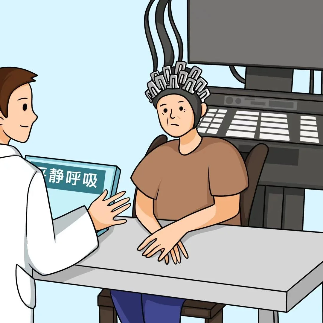
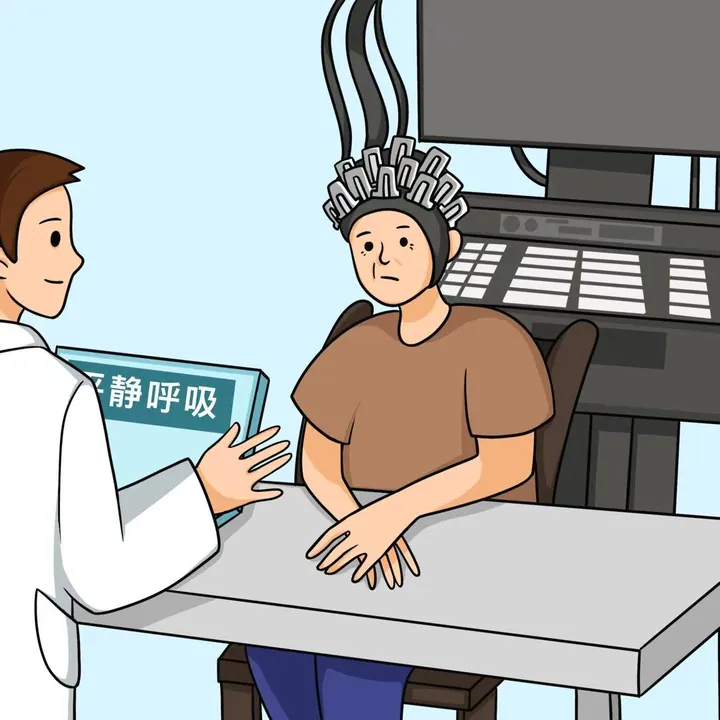
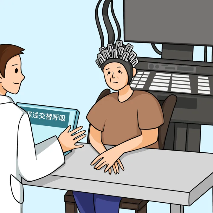
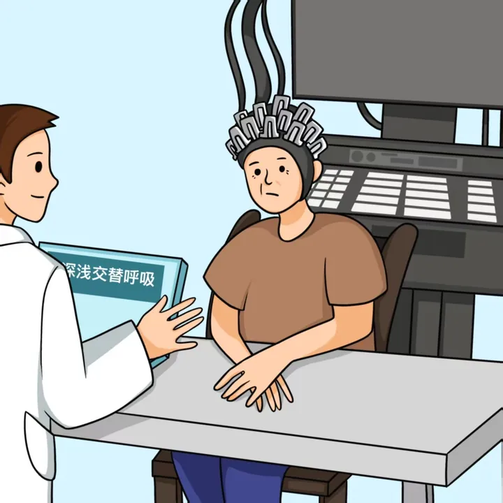
A typical EEG scene: a clinician fitting electrodes while a screen reads "resting state". Such calm, cooperative measurement is foundational to brain research. The filename contains "breath", but that is not enough to tie the image to breathing-pattern research; archived as a resting-state EEG scene.
The Story Behind the Image
Paper abstract (original)
Aims Sleep deprivation (SD) has become a health problem in modern society due to its adverse effects on different aspects. However, the relationship between sleep and cardiovascular system function remains unclear. Here we explored the changes occurring in the brain and the heart sounds after SD. Methods Ninety healthy adult men were recruited and subjected to 36 h of Sleep Deprivation (SD). They participated in a number of tests, including measurements of the heart sound, blood oxygen, and heart rate every 2 h. By using of principal component analysis to reduced the dimensionality of heart sound data. While the ALFF and ReHo indexes were measured via fMRI before and after SD. Correlation and regression analyses were used to reveal the relationship between fMRI and heart sound changes due to SD. Results In this study, there were no abnormal values in the heart rate and blood oxygen during 36 h of SD, whereas the intensity of heart sounds fluctuated significantly increased and decreased. The ALFF was increased in bilateral pericalcarine(Calcarine), left anterior cuneus, (Precuneus_L), right superior temporal gyrus(Temporal_Sup_R), left supplementary motor area (Supp_Motor_Area_L); However, it was reduced in the right medial superior frontal gyrus (Frontal_Sup_Medial_R), right dorsolateral superior frontal gyrus (Frontal_Sup_R) and left medial frontal gyrus (Frontal_Mid_L). The regression analysis uncovered that the intensity of the heart sound in the systole, s1, and s2 phase could be explained by Calcarine_L changes. Conclusion Acute sleep deprivation affects cardiac-brain axis and the specific brain regions. Calcarine_L changes during sleep deprivation are involved in regulating heart contractions.